
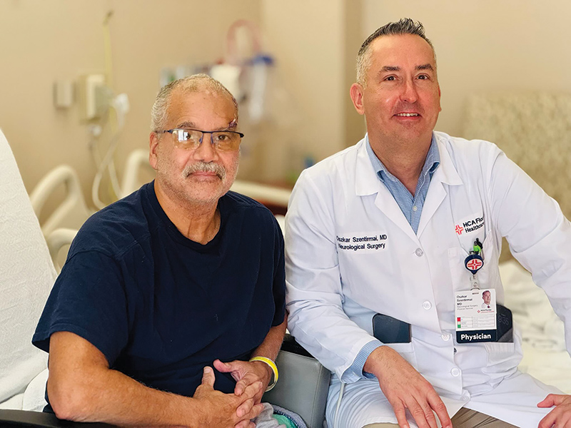
The last thing Francisco Rosado remembers seeing was an apple displayed on a cellphone by the neurosurgeon operating on his brain.
Rosato was awake and talking to the surgical team at the time.
“I said, ‘It’s an apple,’ and then they closed me up,” he recalled.
That moment was the culmination of a highly specialized procedure that allowed neurosurgeon Dr. Oszkar Szentirmai of HCA Florida Lawnwood Hospital to operate near critical areas of Rosado’s brain while continually monitoring his ability to speak and respond.
It also marked a milestone for Lawnwood. Dr. Szentirmai said Rosado’s procedure was the first awake craniotomy performed at the hospital.
“This procedure is typically only done at major academic centers,” Dr. Szentirmai said. “Lawnwood Hospital now has those specialized anesthesiologists and neurosurgical teams in place so that we can safely perform them here on the Treasure Coast.”
For Rosado, the reason for undertaking such an unusual operation was serious. He had been diagnosed with a grade 4 glioblastoma, an aggressive brain tumor located in the left frontal lobe affecting the speech center.
“I chose the awake craniotomy procedure to ensure there would be no deficits affecting speech following surgery because imaging navigation alone is not predictive of post op deficits,” Dr. Szentirmai explained.
The idea of being conscious during brain surgery sounds frightening. Rosato, however, said he took the news calmly when Dr. Szentirmai explained what would be required.
“I guess I have to do it,” he said.
An awake craniotomy is not quite what it sounds like. The patient is not necessarily awake and fully conversant for every minute of the operation. Instead, anesthesia and medication are carefully managed so the patient can be comfortable and responsive when the surgical team needs to test brain function.
The scalp is numbed so the surgeon can open the skull without causing the kind of pain one might imagine from brain surgery. Once the surgeon reaches the brain, the patient can be awakened enough to communicate and perform simple tasks.
And that is where the procedure becomes remarkable.
The brain itself does not have pain receptors, Dr. Szentirmai explained, allowing surgeons to work on brain tissue while the patient is awake. During the operation, the patient may be asked to talk, count, identify pictures, name objects, move fingers or toes, or perform other simple tasks while the surgical team monitors the responses.
The purpose is not to make the surgery more dramatic. It is to make it safer.
Dr. Szentirmai offered a simple analogy: Think of the brain as a city crisscrossed by highways.
Some of those highways carry speech. Others carry signals responsible for movement, sensation or other essential functions. A tumor may be located dangerously close to one of those routes.
The surgeon cannot simply look at an MRI and assume that a particular spot controls a particular function. Brain function varies from person to person, and the surgical team needs to know what is actually happening at the affected location in that patient’s brain.
During an awake craniotomy, the team can stimulate areas of the brain and watch for changes in the patient’s responses. If stimulating a particular area causes the patient to stop speaking, for example, the surgeon has learned that the area or pathway is important to speech. “It identifies a specific pathway,” Dr. Szentirmai said.
The analogy becomes particularly useful when the surgeon encounters the brain’s equivalent of an interstate highway.
“You don’t cross Highway 95,” Dr. Szentirmai said, explaining that some pathways carry vital information between different parts of the brain. Instead, the surgeon looks for a safe route around them.
That is one of the major advantages of having a patient awake and participating in the operation. Rather than relying solely on an MRI image or surgical navigation system to determine where it is safe to operate, the team can continually test the patient’s actual brain function.
For Rosado, that testing involved looking at pictures on Dr. Szentirmai’s cellphone.
Rosado had warned the medical team that he had difficulty distinguishing certain colors, so Dr. Szentirmai took that into account as he showed him images during the procedure. Rosato does not remember the first pictures very well, but he distinctly remembers the last one. An apple.
He identified it correctly, and the surgical team finished the procedure.
Rosado also speaks both English and Spanish, another consideration for the surgical team. Dr. Szentirmai said the anesthesia team was prepared to communicate with him in both languages so that his ability to communicate could be monitored throughout the operation.
Rosado doesn’t remember those conversations. In fact, he remembers very little about the surgery, which lasted about three hours, according to Dr. Szentirmai. Rosado was then transferred to rehabilitation, where he spent about two weeks.
He said that by the day after surgery, things were already beginning to come back together, including his memory and recognition of family members. He has since begun the next phase of treatment, including radiation and chemotherapy.
For Dr. Szentirmai, the objective of surgery is not simply to remove as much tumor as possible. It is to remove as much diseased tissue as possible as safely possible while preserving the abilities that allow a patient to communicate, move, recognize loved ones and maintain quality of life.
That balance is particularly important when a tumor lies close to areas of the brain responsible for essential functions.
Dr. Szentirmai compared treatment of an aggressive brain tumor to a boxing match.
Surgery delivers the first major punch, removing as much of the tumor as possible. The next round involves radiation combined with the oral chemotherapy drug temozolomide, which is used as part of standard treatment for newly diagnosed glioblastoma. Additional cycles of chemotherapy may follow, along with regular imaging to monitor the patient.
If the tumor recurs, additional treatment options may include another surgery, radiation, chemotherapy or other therapies depending on the individual patient and the biology of the tumor. “This is where research comes into play,” Dr. Szentirmai said, explaining that studying a patient’s tumor tissue may eventually help physicians better understand which treatments are most likely to work against that particular cancer.
The goal, he said, is increasingly personalized treatment based on the biology of an individual patient’s tumor rather than relying on a one-size-fits-all approach.
But before any of those treatments can begin, the first challenge is getting as much tumor as possible while protecting the brain around it.
“Rosado’s speech difficulty, memory, balance and weakness problems all resolved working two to three weeks after surgery, so we achieved the goal to cause no harm. His speech is actually better after brain tumor surgery,” Dr. Szentirmai said.
For Rosado, however, the most remarkable part of his surgery may be what he doesn’t remember.
He doesn’t remember the drilling or the surgical instruments. He doesn’t remember a lengthy conversation with his surgeon. He doesn’t remember being frightened.
He remembers a picture of an apple.
For a neurosurgeon, the patient’s recognition of a piece of fruit represented something extraordinary – a demonstration that Rosado’s brain was still communicating with the outside world while the surgeon worked inside his skull.
Dr. Oszkar Szentirmai’s office is located at HCA Florida Neurosurgical Specialists-Lawnwood, 2401 Frist Boulevard, Suite 6, Fort Pierce. Call 772-241-6720 for an appointment.